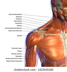
Shoulder Muscles Diagram / Shoulder Muscles And Chest Human Anatomy Diagram Am Medicine Shoulder Muscle Anatomy Human Body Anatomy Muscle Diagram / Although three ligaments protect and surround the shoulder joint, most of its stability comes from the powerful muscles and tendons of the rotator cuff.
byMargo Padilla-
0
Shoulder Muscles Diagram / Shoulder Muscles And Chest Human Anatomy Diagram Am Medicine Shoulder Muscle Anatomy Human Body Anatomy Muscle Diagram / Although three ligaments protect and surround the shoulder joint, most of its stability comes from the powerful muscles and tendons of the rotator cuff.. 6 photos of the shoulder muscles labelled diagram. Diagram shoulder muscles human anatomy shoulder muscles amazing neck and shoulder muscles. The primary function of the shoulder girdle is to give strength and range of motion to the arm. Related online courses on physioplus. The shoulder muscles are a set of complex muscles that act as a link between the torso and the head or neck.
Shoulder flexion is movement of the shoulder in a forward motion. The shoulder muscles produce the characteristic shape of the shoulder and can be classified into two groups: Learn vocabulary, terms and more with flashcards, games and other study tools. The core muscles are those in the abdomen, back, and pelvis, and they. Groin muscles diagram diagram of groin aponeurosis from sscsantry groin project medical.
Shoulder Anatomy Labeled Hd Stock Images Shutterstock from image.shutterstock.com There are three main muscles in your shoulder: It is the major joint connecting the upper limb to the trunk. The shoulder blades, which are prominent unless the back muscles are so developed they cover them up. Related online courses on physioplus. The two large main muscles of this. Terms in this set (81). An example of shoulder flexion can be seen when reaching forward to grasp an object. Neck and shoulder muscles diagram.
Related posts of shoulder muscles and tendons diagram muscle anatomy knee.
The anterior deltoid, the lateral deltoid, and the posterior deltoid. Diagram shoulder muscles human anatomy shoulder muscles amazing neck and shoulder muscles. The shoulder muscle tissues can be readily injured and therefore being aware of the appropriate strategy is pretty significant when functioning out. Other muscles that aid in shoulder movement include: Shoulder muscles, pictures and descriptions of the movements and attachments. This diagram with labels depicts and explains the details of. Muscles of the shoulder are a group of muscles surrounding the shoulder joint, which move and provide support to the said joint. The two large main muscles of this. There are three main muscles in your shoulder: The primary function of the shoulder girdle is to give strength and range of motion to the arm. From the arm muscle diagram above, the muscles of the arm that can be seen easily on the surface include biceps, triceps, brachioradialis, extensor. The shoulder muscles produce the characteristic shape of the shoulder and can be classified into two groups: Supraspinatus, infraspinatus, ters minor,.et), using interactive animations and labeled diagrams.
Terms in this set (81). This rotator cuff muscle helps with the raising and lowering of the upper arm. Printable shoulder muscles diagrams to help you study the muscles structure in human's shoulder. The teres minor, subscapularis, supraspinatus, and infraspinatus muscles together form the rotator cuff, which stabilizes the humeral head (the ball. The visibility of the shoulder blades also varies note also less bulky shoulders and a waist that's less thin.
Muscles Of The Human Body Art Rocket from www.clipstudio.net The anterior deltoid, the lateral deltoid, and the posterior deltoid. See below to view an image of the rotator cuff structure: The shoulder muscles bridge the transitions from the torso into the head/neck area and into the upper extremities of the arms and hands. The shoulder muscles are a set of complex muscles that act as a link between the torso and the head or neck. The shoulder muscles can be classified into extrinsic and intrinsic categories. Diagram shoulder muscles human anatomy shoulder muscles amazing neck and shoulder muscles. This rotator cuff muscle helps with the raising and lowering of the upper arm. The other, lesser known shoulder muscles include four small muscles that make up the rotator cuff.
The human shoulder is made up of three bones:
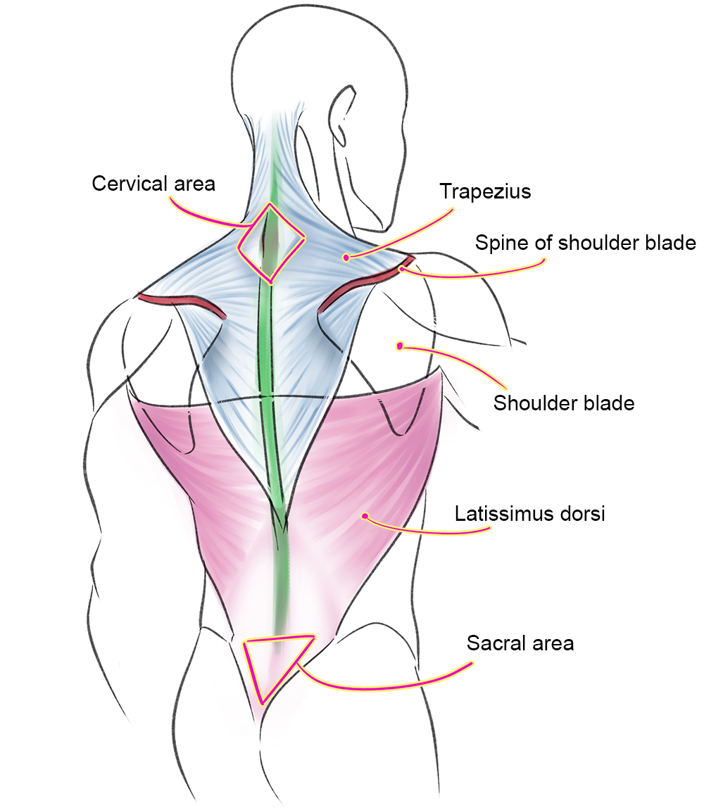
The extrinsic muscles of the shoulder include trapezius, latissimus dorsi, levator scapulae, rhomboid major and rhomboid minor. This rotator cuff muscle helps with the raising and lowering of the upper arm. The shoulder muscles are a set of complex muscles that act as a link between the torso and the head or neck. The visibility of the shoulder blades also varies note also less bulky shoulders and a waist that's less thin. Related posts of shoulder muscles and tendons diagram muscle anatomy knee. An example of shoulder flexion can be seen when reaching forward to grasp an object. Printable shoulder muscles diagrams to help you study the muscles structure in human's shoulder. Shoulder flexion is movement of the shoulder in a forward motion. This diagram with labels depicts and explains the details of. Diagram shoulder muscles human anatomy shoulder muscles amazing neck and shoulder muscles. Diagram muscle shoulder joint (page 1) 2. Shoulder muscle and ligament diagram. Sternum shoulder muscles **muscles on anterior aspect pec.
Human anatomy diagrams show internal organs, cells, systems, conditions, symptoms and sickness information and/or tips for healthy living. Just like the muscle tissues in unique elements of the human physique, even our shoulder muscle tissues are prone to standard put on and tear. Shoulder muscle and ligament diagram. The rotator cuff is a complex and delicate structure of. This diagram with labels depicts and explains the details of.
Human Muscle Diagram Graph Diagram from graphdiagram.com Sternum shoulder muscles **muscles on anterior aspect pec. Although three ligaments protect and surround the shoulder joint, most of its stability comes from the powerful muscles and tendons of the rotator cuff. The muscular system consists of various types of muscle that each play a crucial role in the function of the body. Shoulder muscle and ligament diagram. This diagram depicts shoulder muscle diagram. Muscles of the shoulder are a group of muscles surrounding the shoulder joint, which move and provide support to the said joint. Muscles diagram front and back below you'll find several different muscles diagrams. Related online courses on physioplus.
6 photos of the shoulder muscles labelled diagram.
Muscles allow a person to move muscle tendons in the knee joint and the shoulder joint are crucial in stabilization. Although three ligaments protect and surround the shoulder joint, most of its stability comes from the powerful muscles and tendons of the rotator cuff. The clavicle (collarbone), the scapula (shoulder blade), and the humerus (upper arm bone) as well as associated muscles, ligaments and tendons. Related online courses on physioplus. This goes for females as well, except that their pectoral muscles are hidden behind the. The rotator cuff is a complex and delicate structure of. The shoulder muscles produce the characteristic shape of the shoulder and can be classified into two groups: This rotator cuff muscle helps with the raising and lowering of the upper arm. Shoulder joint of human body anatomy infographic diagram with all parts including bones ligaments muscles bursa cavity capsule cartilage membrane for medical science education and health care. See below to view an image of the rotator cuff structure: Shoulder flexion is movement of the shoulder in a forward motion. The muscular system consists of various types of muscle that each play a crucial role in the function of the body. Human anatomy and physiology diagrams: